What and where are the cruciate ligaments?
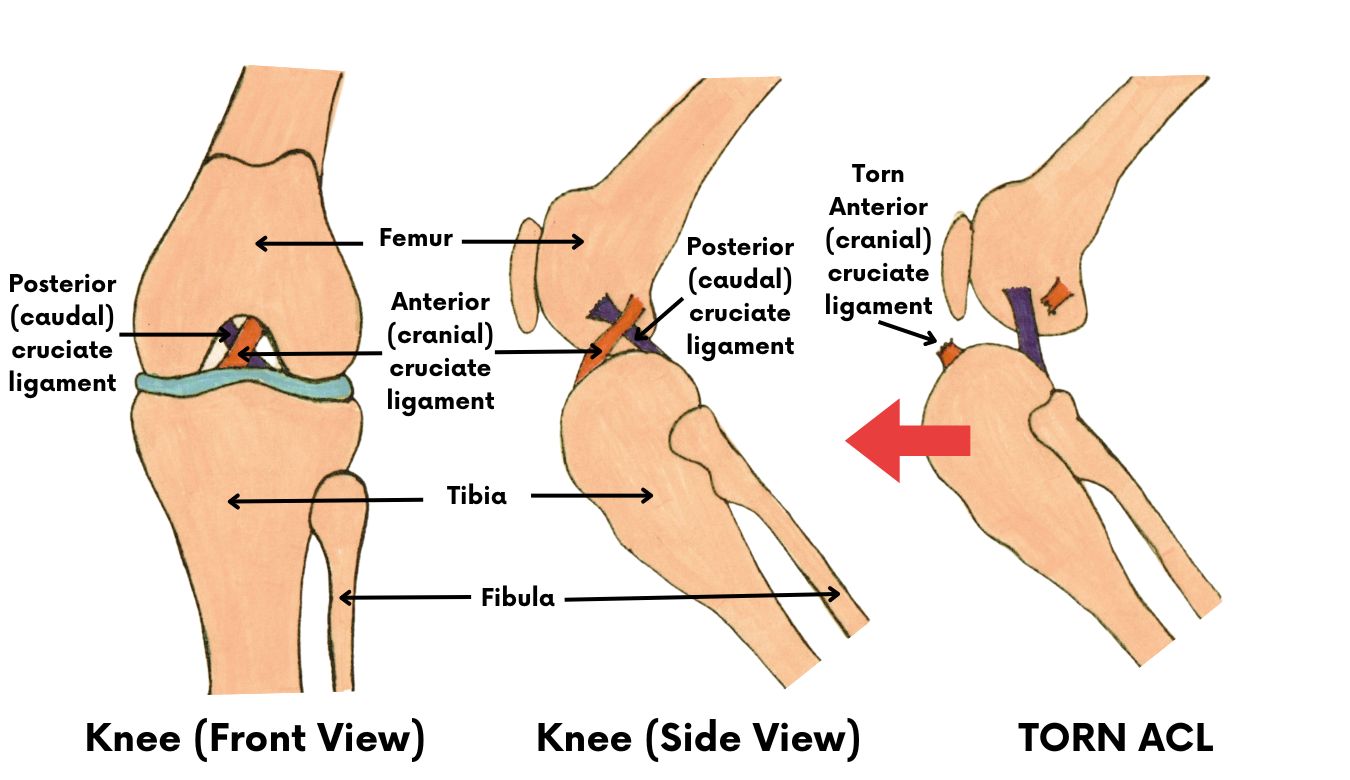
There are two bands of fibrous tissue called the cruciate ligaments in each knee joint. They join the femur and tibia (bones above and below the knee) together so that the knee works as a hinged joint.
They are called cruciate ligaments because they “cross over” inside the knee joint. One ligament connects from inside to outside the knee joint and the other outside to inside, crossing each other in the middle.
Humans have the same anatomical structure of the knee. Cruciate ligament rupture is a common knee injury of athletes.
How does the injury occur?
The knee joint is a hinged joint and only moves in one plane, backwards and forwards. Traumatic cruciate damage is caused by a twisting injury to the joint. This is most often seen in dogs and athletes when running and suddenly changing direction so that the majority of the weight is taken on this single joint. This injury usually affects the anterior (front) ligament. The joint is then unstable and causes extreme pain, often resulting in lameness.
The injury also occurs commonly in obese dogs, just by stumbling over a pebble while walking.
A more chronic form of cruciate damage can occur due to weakening of the ligaments as a result of disease. The ligament may become stretched or partially torn and lameness may be only slight and intermittent. With continued use of the joint, the condition gradually gets worse until rupture occurs. Approximately 33% of dogs will tear the cruciate ligament of the opposite limb.
Is an operation always necessary?
Dogs under 10 kgs (22 lbs.) may heal without surgery. These patients are often restricted to cage rest for two to six weeks. Dogs over 10 kg. (22 lbs.) may require surgery to heal. Unfortunately, most dogs will eventually require surgery to correct this painful injury.
What surgical options are available at Belton Veterinary Clinic?
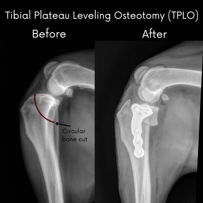
Tibial Plateau Leveling Osteotomy (TPLO)
TPLO levels the slope of the tibia by cutting the bone and rotating it. A plate is fastened onto the side of the bone with a set of screws to hold the tibia together during the healing process. The TPLO keeps the femur bone from shifting backward during weight-bearing activities and allows other supporting structures of the stifle to stabilize the joint.
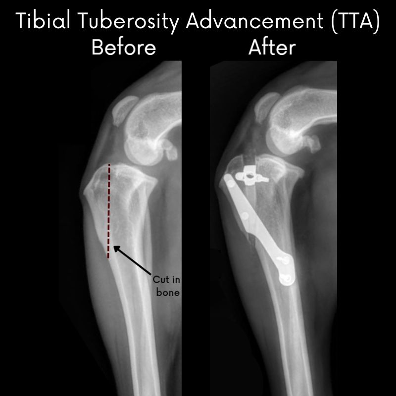
Tibial Tuberosity Advancement (TTA)
The TTA procedure  involves making a cut in the front part of the tibia bone (tibial tuberosity) and advancing this portion of bone forward in order to realign the patellar ligament so that the abnormal sliding movement within the knee joint is eliminated. A specialized bone spacer, plate and screws are used to secure the bone in place.
- Your dog’s leg may look different because the tibial crest protrudes more after surgery.
- The dog may walk with the stifle in a more flexed angle.
Benefits of TPLO and TTA:
- Faster recovery
- Earlier use of the limb after surgery
- Better chance to return to full activity
- Better range of motion of the joint
About half of all canine patients will begin walking on the injured leg within 24 hours after surgery.
At 2 weeks postoperatively, most dogs are bearing moderate to complete amounts of weight on the affected leg.
By 10 weeks, most dogs do not have an appreciable limp or gait abnormality.
As mentioned above, at 4 months postoperatively, the majority of dogs can begin walking and playing normally, with only the most stressful activities restricted.
Within 6 months, most dogs can resume full physical activity.
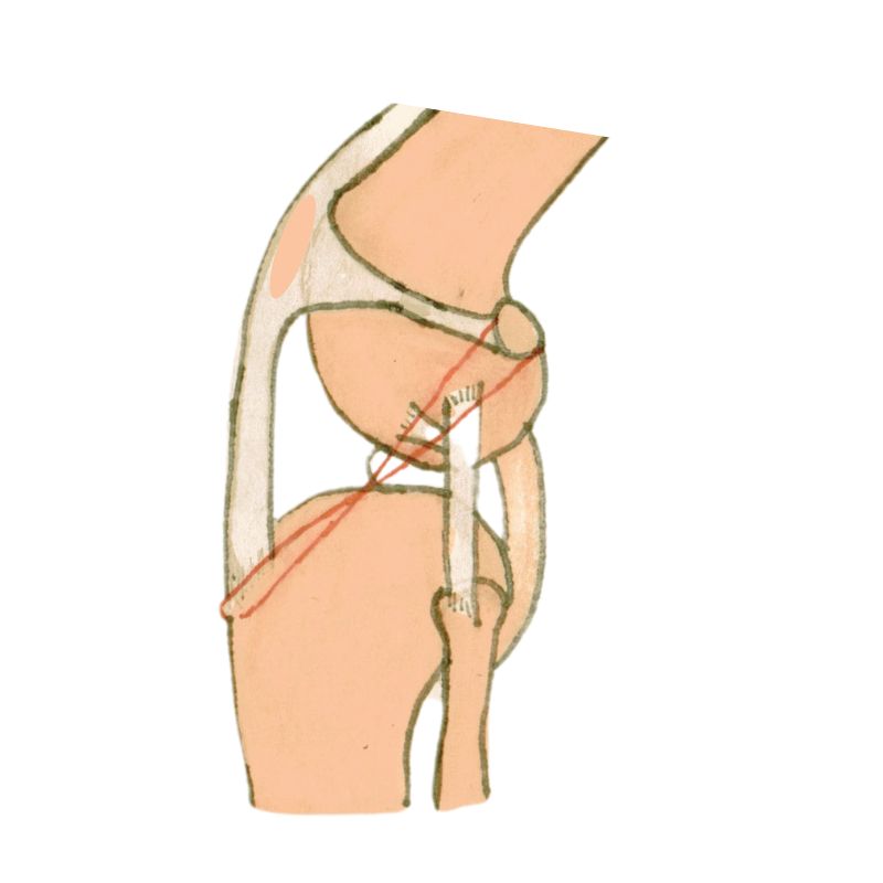
Lateral Fabellar Suture (Extracapsular Repair)
This procedure involves using medical grade nylon that is implanted in the leg to mimic the action of the cranial cruciate ligament. It will decrease tibial thrust and internal rotation. Over time, scar tissue will form to help stabilize the joint.
- 85% of the cases are significantly improved from their preoperative state.
- About 50% of these dogs will have some degree of lameness, whether it is mild or intermittent following heavy activity.
- Even though this surgery may not return the limb to perfectly normal function, these dogs usually are greatly improved over their condition prior to surgery.
- The lateral fabellar suture technique will not stop the progression of arthritis that is already present in the joint. As a result, your pet may have some stiffness of the limb in the mornings. In addition, your pet may have some lameness after heavy exercise or during weather changes. To help with stiffness chondroitin sulfate and glucosamine may be given.
We can recommend which procedure is best for your dog. This is usually based on weight, age and activity level. The recovery period is the most important part. It is imperative to follow the post-op guidelines. There should be no off leash activity until the veterinarian gives you the ok. This is usually 8 weeks.